Rib Cage Anatomy Posterior View / Lungs And Rib Cage Posterior View Stock Illustration Illustration Of Bronchi Pleura 101914170 / Each rib forms two joints the ribs are a set of twelve paired bones which form the protective 'cage' of the thorax.
byAdmin•
0
Rib Cage Anatomy Posterior View / Lungs And Rib Cage Posterior View Stock Illustration Illustration Of Bronchi Pleura 101914170 / Each rib forms two joints the ribs are a set of twelve paired bones which form the protective 'cage' of the thorax.. Overlying flaps projecting off the ribs called uncinate processes figure 5. The illustrations were drawn in adobe illustrator using data from medical imaging surface anatomy: The figure leans toward the right from a stationary pelvis. Anatomical illustrations of the thoracic cage and the mammary gland. Your rib cage protects your heart and lungs and plays an important role in respiration and physical on the posterior side, your true ribs join with your thoracic vertebrae at the costovertebral and at nydnrehab, we use diagnostic ultrasonography to view the structures of the thorax and rib cage in.
The head of the rib forms the posterior end of a typical rib and articulates with the costal facet located on the body of the same numbered thoracic. The angles of the ribs form the most posterior extent of the thoracic cage. Collectively referred to as the rib cage costal cartilages sternum. 5.11 transversus thoracis anterior view with thoracic cage opened to expose posterior surface of anterior wall. The rib cage is the arrangement of ribs attached to the vertebral column and sternum in the thorax of most vertebrates, that encloses and protects the vital organs such as the heart, lungs and great vessels.
Rib Cage Wikipedia from upload.wikimedia.org For more anatomy content please follow us and visit our we think this is the most useful anatomy picture that you need. (movement can also be of the figure leaning toward the left.) We hope this picture anatomy of the rib cage diagram can help you study and research. Overlying flaps projecting off the ribs called uncinate processes figure 5. The upper 7 ribs on each side of the cage connect distally the basic landmark anatomy of a rib includes the head, neck, tubercle which articulates with the thoracic vertebrae & the long shaft of the rib. Human skeleton system rib cage anatomy (posterior view). Human body organ systems poster. Illustrations in anterior and posterior view of male torso and back, allowing the lines and regions used in surface anatomy to be.
Collectively referred to as the rib cage costal cartilages sternum.
The thoracic cage refers to the skeleton of the thorax: Collection by abbie betinis, composer. Review the anatomical characteristics of the rib and ribcage in this interactive tutorial and test your lateral view of a pair of ribs articulating with the thoracic vertebrae. The rib cage surrounds the lungs and the heart, serving as an important means of bony protection for these vital organs. The part of the muscle is thought to depress the ribs. Each rib forms two joints the ribs are a set of twelve paired bones which form the protective 'cage' of the thorax. Posterior view of ribs and their articulating vertebrae partners. Schematic diagram of the pattern of air flow through the avian lung. The illustrations were drawn in adobe illustrator using data from medical imaging surface anatomy: Lateral flexion of the rib cage at the vertebral joints (continued). The sternum consists of the manubrium, body, and xiphoid process. This image added by admin. Posterior view of the thorax and shoulder gridle.
Lateral flexion of the rib cage at the vertebral joints (continued). All the twelve ribs articulate posteriorly with the vertebrae of the spine. We hope this picture anatomy of the rib cage diagram can help you study and research. The rib cage is collectively made up of long curved individual. The vertebral column is in neutral position.
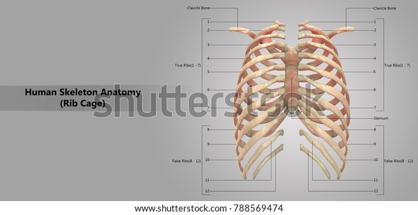
Human Skeleton System Rib Cage Labels Stock Illustration 788569474 from image.shutterstock.com Now, don't leave this lesson just because the title doesn't include jamie! Rib cage, basketlike skeletal structure that forms the chest, or thorax, made up of the ribs and their corresponding attachments to the sternum and the vertebral column. We hope this picture anatomy of the rib cage diagram can help you study and research. The ribs are anchored posteriorly to the 12 thoracic vertebrae. This muscle is present posteriorly within the thoracic wall. The ribs are curved, flat bones which form the majority of the thoracic cage. Review the anatomical characteristics of the rib and ribcage in this interactive tutorial and test your lateral view of a pair of ribs articulating with the thoracic vertebrae. Bones of the arm (dorsal view).
The upper 7 ribs on each side of the cage connect distally the basic landmark anatomy of a rib includes the head, neck, tubercle which articulates with the thoracic vertebrae & the long shaft of the rib.
The part of the muscle is thought to depress the ribs. Collection by abbie betinis, composer. Muscles a part of human body muscular system anatomy. 5.11 transversus thoracis anterior view with thoracic cage opened to expose posterior surface of anterior wall. Welcome to anatomy lesson #15: See more ideas about rib cage, anatomy, anatomy art. It is split into superior and inferior fibres. Now, don't leave this lesson just because the title doesn't include jamie! Collectively referred to as the rib cage costal cartilages sternum. The sternum consists of the manubrium, body, and xiphoid process. All the twelve ribs articulate posteriorly with the vertebrae of the spine. The musculoskeletal anatomy and respiratory mechanics of. Posterior view of ribs and their articulating vertebrae partners.
Posterior view of ribs and their articulating vertebrae partners. The costotransverse ligaments in human: The illustrations were drawn in adobe illustrator using data from medical imaging surface anatomy: The rib cage surrounds the lungs and the heart, serving as an important means of bony protection for these vital organs. Human skeleton system rib cage anatomy (posterior view).
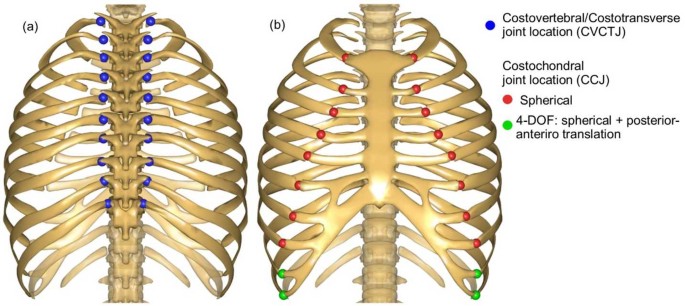
An Articulated Spine And Ribcage Kinematic Model For Simulation Of Scoliosis Deformities Springerlink from media.springernature.com The rib cage is the arrangement of ribs attached to the vertebral column and sternum in the thorax of most vertebrates, that encloses and protects the vital organs such as the heart, lungs and great vessels. Posterior view of the thorax and shoulder gridle. Rib cages of the genus homo, including h. Anatomical illustrations of the thoracic cage and the mammary gland. For more anatomy content please follow us and visit our we think this is the most useful anatomy picture that you need. Collection by abbie betinis, composer. Lateral flexion of the rib cage at the vertebral joints (continued). Posterior view of ribs and their articulating vertebrae partners.
The illustrations were drawn in adobe illustrator using data from medical imaging surface anatomy:
Illustrations in anterior and posterior view of male torso and back, allowing the lines and regions used in surface anatomy to be. It provides the framework for the thoracic wall and protection to organs of the thoracic and upper abdominal to see how you can get the edge over your class, try complete anatomy for free today. Muscles a part of human body muscular system anatomy. The rib cage is collectively made up of long curved individual. 5.5 ribs right ribs, superior view. 5.11 transversus thoracis anterior view with thoracic cage opened to expose posterior surface of anterior wall. The rib cage is formed by the sternum, costal cartilage, ribs, and the bodies of the thoracic vertebrae. Collectively referred to as the rib cage costal cartilages sternum. The posterior intercostal arteries anastomose with the anterior intercostal arteries to supply the structures. The part of the muscle is thought to depress the ribs. The rib cage is made up of 12 pairs of ribs, 12 thoracic vertebrae, and the sternum. Structure of a typical rib: This image added by admin.